[CIRP note: CAUTION: This important paper from 1933 does not contain current recommendations for the care of the intact penis. Dr. Deibert's discussion of retraction on the eighth day does NOT represent current practice. Forcible premature retraction may injure the child. Retraction of the prepuce should be delayed until the child can do it for himself.]
THE SEPARATION OF THE PREPUCE IN THE
HUMAN PENIS
Glenn A. Deibert
The Daniel Baugh Institute of Anatomy, Jefferson Medical College
One text figure and three plates (nine figures)
As is well known, the prepuce in the human penis is adherent to the glans at birth, a layer of stratified squamous epithelium being interposed, and shortly after birth these parts become separated so that the prepuce may be retracted. Failure of the prepuce to separate from the glans spontaneously or neglect to separate the parts by artificial means results in phimosis or inability to retract the prepuce. Frequently, the customary retraction about the eighth day of infancy exposes irregularly distributed bleeding points, indicating incomplete separation.
It is the purpose of this study to ascertain more definitely the nature of this cleaving process, the time of its onset, and the age when ultimate separation is completed.
LITERATURE
Numerous papers dealing primarily with certain other phases of the embryology of the human penis make only secondary reference to the mechanism of preputial separation. For the most part these observations are based on one or at best few age periods only and usually refer to material before birth. In no instance is the process followed from beginning to end in a serial study.
Bokai (1860) was the first to direct attention to the physiological adherence of the foreskin. Schweiger and Seidel (1866) gave the first description of the development of the prepuce in the human. In the later stages of foetal development, they noted the appearance of `concentric masses' of cells within the epithelium intervening between the glans and the prepuce. The centers of these masses were found to break down and the resulting cavities to coalesce to effect a separation. These observations were confirmed by Tourneux (1877), Retterer (1890), Hart ('07), Fleischman ('07), Jones ('10), and Johnson ('22). Edington ('10) noted clinically in circumcision that spontaneous separation does not always progress to the same degree in children of the same age. In many instances he found considerable bleeding on retracting the prepuce. The consistent finding of a free prepuce for a variable distance in the region of the meatus and in the area back of the corona, designated as the sulcus in the adult, led Edlington to think that separation begins anteriorly and posteriorly and processes toward the center.
MATERIALS
A series of twenty-nine penes from infants, with ages ranging from 6 months prematurity to six months after birth were prepared and studied. Of this number, fourteen were taken from stillborn infants and eleven from negro babies. The specimens were fixed in Bouin's fluid, blocked in paraffin and sectioned serially at 10 mu. Every fourth section was mounted and stained with haemotoxylin and eosin.
EMBRYOLOGY
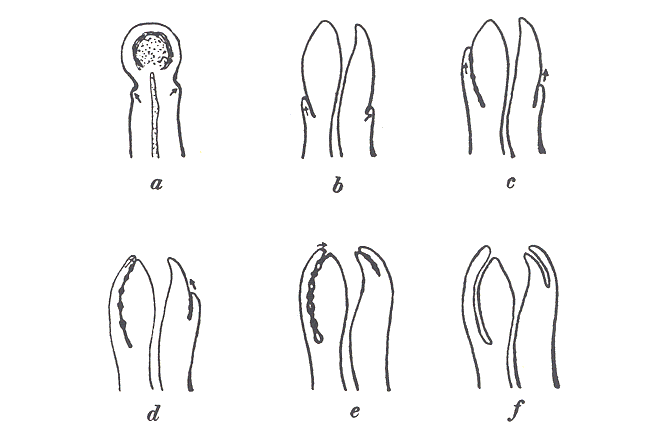
It is necessary to recall the development of the prepuce briefly so that its subsequent separation may be followed to better advantage. The prepuce is often incorrectly described as growing forward as a free unattached sheath. In a 75-day foetus, the end of the penis is not covered by a prepuce but by a thick epidermal layer continuous with the anterior body wall. The distal end of the penis, or phallus, as it is referred to at this age, represents the glans, but no prepuce is in evidence (Fig. 1A). In a 55-mm. embryo, the glans is delimited from the shaft on its dorsal and lateral walls by a constriction. Here the epithelium present a semicircular thickening, the glandular lamella of Fleischman, which projects into the mesenchyme of the penis (Fig. 1 B). Gradually this point of origin of the glandar lamella shifts toward the tip, so that in general direction in sagittal section is no longer transverse but longitudinal. The deep or free margin does not change from it original position so that the glandar lamella becomes curved. This curve marks the corona of the glans. The shifting continues, so that in a 170-mm. embryo the attached margin of the glandar lamella is found at the external urinary meatus, thereby completing the development (fig. 1 D and E). The lamella thickens during the shifting process, epithelial pearls appear and the process of separation is inaugurated.

Fig. 1 Outline drawings showing the
progressive stages
in the development and separation of the
prepuce.
OBSERVATIONS ON THE PROCESS OF SEPARATION
The youngest penis studied was taken from a 6-month premature foetus. This already shows evidence of a beginning separation. The glandar lamella is well defined, terminating posteriorly in a bulb-like enlargement at the site of the sulcus in the adult penis. At various levels, a large, single, concentric epithelial mass or epithelial pearl occupies this enlargement (fig. 2). The center of the pearl shows a densely eosin-staining laminated mass of keratin, an evidence of early degeneration. Anterior to the enlargement, numerous nodular thickenings occur along the course of the lamella. In these thickenings, the cells are again arranged in whorl-like fashion, but these pearls are of much smaller size and show no central degenerative changes. Their incidence varies with the level of the section. At one level eighteen pearls were observed in one arm of the lamella and seven in the other. Anteriorly, where the glandar lamella joins the epidermis, cellular changes also indicate some separation (fig. 4). There is a ragged indentation at the junction filled with loose strands of hyalinized squamous cells continuous with the outer layers of the epidermis of the prepuce. It is clear, then, that the separative process begins earlier than the 6-month stage of foetal development.
The cellular makeup of the larger pearls in the posterior enlargement is of interest. The layers of cells of these pearls correspond to the layers of the epidermis (fig. 3). The peripheral layer of the glandar lamella corresponds to the basal cell of the stratum germinativum. Its cells are columnar in shape; its nuclei spherical and prominent. Within this basal cell layer, the cells are more rounded or polyhedral in shape; its nuclei spherical and prominent. Within this basal cell layer, the cells are more rounded or polyhedral in shape -- the prickle cell layer. The intercellular bridges are especially prominent. As the pearl is approached the cells gradually assume a flat shape, the cytoplasm becomes granular and the nuclei spindle-shaped, simulating the stratum germinativum of the skin. Immediately surrounding the hyalinized center of a pearl are several layers of very flat cells with prominent eleidin granules, resembling the stratum granulosum of the skin. The periphereal portion of the hyalinized center is made up of several layers of squamous cells whose cytoplasm is distinctly hyaline and whose nuclei are pyknotic. The central part of the hyalinized center is laminated and shows few fragments of disintegrated nuclei. It corresponds to the horny layer of the epidermis.
In the stillborn specimens varying degrees of pearl formation and separation are present. In the smaller specimens the pearls are very numerous but little keratinization is found in their centers (fig. 8). All of these smaller specimens exhibit also some degree of anterior separation, cornified material lining the break, which is significant of keratinization. In several specimens, the anterior separation extends posteriorly half the length of the glandular lamella. In the larger stillborn specimens, the pearls are larger and more numerous. In many instances their centers show extensive keratinization (fig. 2). The loose arrangement of the keratin resembles the laminations of a Meisner's corpuscle. The anterior separation is pronounced in most instances.
In two of the larger stillborn specimens and in one from a child which had lived several hours, a large specimen also, the pearls are elongated and almost approximate each other. The day-old penis shows even more extensive separation. Along one arm of the lamella the prepuce is completely free at all levels studied. The other arm shows an anterior separation for some distance (fig. 7). The pearls along the course of the lamella are again elongated and almost approximate one another. Their centers show keratization. Between the pearls the cells are flattened longitudinally and show a granular cytoplasm. In some sections the cells appear hyalinized. Apparently, this is an interpearl keratization, serving to link the degenerated centers of the pearls into a continuous separation. (fig. 6).
The 10-day penis shows almost complete separation (fig. 9). Small areas of adhesions persist posteriorally in both arms at various levels. A free, thick stratum of keratin occupies the interval between the separated prepuce and glans. In the remaining older specimens, varying degrees of adhesions persist, even a slight amount at the age of 4 and 6 months (fig. 10). These adhesions are confined to a small portion of the posterior third of the glandar lamella. In this persisting area, degenerating pearls are again in evidence.
SUMMARY
The separation of the prepuce is the human penis is essentially a process of keratization of the intervening epithelium. It begins anteriorly and posteriorly at about the same time and proceeds toward the center. When confined on all sides the separation manifests itself as an epithelial pearl formation. On the surface, as is possible in the anterior region, it appears as a desquamation.
Pearl formation is first seen in the enlarged posterior extremity of the glandar lamella, the site of the sulcus in the adult, approximately the sixth month of foetal life. Later pearl appear at intervals anteriorly along the course of the lamella. At first they are mere arrangements of cells in whorl-like fashion, but later their centers keratinize and leave a cavity. To effect a continuous separation of the prepuce, these cavities may actually coalesce or they may be united by a longitudinal keratinization of the epithelium intervening between the pearls.
Separation is not completed at birth, but is accomplished sometime during infancy or early childhood. Unless the prepuce has been retracted, slight adhesions may persist in the posterior regions of the glandar lamella at 5 and 6 months.
Separation is sufficient at the 10-day stage to allow mechanical retraction without danger of a tear, apparently an important factor in completing the division.
Nutrition or size of the infant is a definite factor in the advancement of the process. The larger specimens at birth show more separation than the smaller ones.
No degree of difference is apparent in the character and rate of separation in the white and negro races.
The author desires to acknowledge his indebtness to Drs. J. Parsons Schaeffer, H. E. Radasch, and H. E. Stewart, and Prof. C. A. Horn, for their assistance and suggestions in the preparation of this work.
                               PLATE 1

                       Explanation of figures

   2  A sagittal section of the penis at term showing the separation
   of the prepuce anteriorally and posteriorly.  Note the thickenings
   of the epithelium in the intermediate segment.

   3  An epithelial pearl, an enlargement of region `a' in figure 2.
   See text, page 390.

   4. Area of desquamation of epithelial cells in the anterior region,
   representing region `b' in figure 2.
|

                               PLATE 2

                       Explanation of figures

   5  A cross section of the penis just before term.  Note the
   numerous areas of separation by the formation of epithelial pearls.

   6  An enlargement of region `a' in figure 7. Note the interpearl
   area of keratinization of the epithelium.  See text, page 391.

   7  A sagittal section of a penis, aged 1 day.  Note the advanced
   stage of the separation of the prepuce on the right side;  the
   presence of a number of epithelial pearls, with less separation of
   the prepuce on the left side.
|

                               PLATE 3

                       Explanation of figures

   8  A sagittal section of the penis near term.  Note that there is
   little real separation of the prepuce in this specimen.

   9. A sagittal section of the penis aged 10 days.  Note the almost
   total separation of the prepuce with a considerable amount of
   keratinized epithelium in the form of debris.

   10  A sagittal section of the penis of an infant aged 4 months.
   Note the advanced stage of the separation of the prepuce on the
   right side.  Posteriorally, there is still an area of adherence.  A
   degenerated epithelial pearl appears on this side.  On the left
   side the prepuce is still largely adherent despite the advanced age
   of the specimen.
|
